Article

Focused Ultrasound in the Emergency Department for Patients with Acute Heart Failure

Register or Login to View PDF Permissions
Permissions× For commercial reprint enquiries please contact Springer Healthcare: ReprintsWarehouse@springernature.com.

For permissions and non-commercial reprint enquiries, please visit Copyright.com to start a request.

For author reprints, please email rob.barclay@radcliffe-group.com.
Average (ratings)
No ratings
Your rating

Abstract

The emergency department (ED) plays a key role in the initial diagnosis and management of acute heart failure (AHF). Despite the advent of novel biomarkers and traditional methods of assessment, such as history, examination, and chest X-ray, diagnosis of the dyspnoeic ED patient is, at times, very challenging. Focused cardiac and pulmonary ultrasound has emerged as a valid, facile and efficient method to aid in the initial diagnosis and management of AHF.

Disclosure:P Pang is or has been in the past year a consultant for Intersection Medical, INSYS, Janssen, Medtronic, Novartis, Trevena, scPharmaceuticals, Cardioxyl, Roche Diagnostics and Relypsa; and has received honoraria from Palatin Technologies. Both M Rutz and FM Russell have no disclosures.

Received:

Accepted:

Correspondence Details:Peter S Pang, 720 Eskenazi Avenue, FOB 3rd Floor, Department of Emergency Medicine, Indiana University School of Medicine, Indianapolis, IN 46032, USA. E: ppang@iu.edu

Copyright Statement:

The copyright in this work belongs to Radcliffe Medical Media. Only articles clearly marked with the CC BY-NC logo are published with the Creative Commons by Attribution Licence. The CC BY-NC option was not available for Radcliffe journals before 1 January 2019. Articles marked ‘Open Access’ but not marked ‘CC BY-NC’ are made freely accessible at the time of publication but are subject to standard copyright law regarding reproduction and distribution. Permission is required for reuse of this content.

The emergency department (ED) plays a critical role in the initial diagnosis and management of acute heart failure (AHF),1,2 as nearly 80 % of all AHF admissions originate from the ER.3 However, patients do not present with a diagnosis; rather they present with a chief complaint reflecting signs and symptoms – most commonly breathlessness. Differentiating AHF from other causes of breathlessness can be challenging, especially in patients with multiple co-morbid conditions such as chronic heart failure and chronic obstructive pulmonary disease (COPD). Failure to diagnose correctly leads to delays or mistreatment, potentially leading to worsened outcomes. Determining whether breathlessness is cardiac or non-cardiac in origin is a critical step in management.

In this article, the authors review the role of focused cardiac and pulmonary ultrasound in the diagnosis and management of dyspnoeic emergency patients, with a specific focus on AHF. Importantly, point- of-care cardiac ultrasound does not replace formal echocardiography (ECG).4,5 As the training requirements for focused ultrasound are beyond the scope of this review, readers should refer elsewhere.6,7

Clinical Case Study

A 68-year-old man presents to the ER complaining of shortness of breath for the past three days, progressively worsening. He has a low-grade fever and his initial vital signs are as follows: HR 109, BP 158/81, RR 20, Sa02 97 % on room air. He has a history of coronary artery disease with two stents placed two years ago. He also has a long history of smoking and has COPD. He has no known history of heart failure but does report increase dyspnoea on exertion, but denies paroxymal nocturnal dyspnoea and orthopnoea. On examination, he is in mild distress, but able to speak in complete sentences. His heart is regular, but fast with no murmurs, gallops or rubs. Faint expiratory wheezes bilaterally are audible. He is mildly obese and jugular venous distention is not ascertained, despite hepatic pressure. He has trace pitting oedema bilaterally. Electrocardiogram (ECG) shows sinus tachycardia, but no acute ischaemic changes.

Background

AHF is a clinical diagnosis. Similar to other diagnostic studies, such as chest radiography and biomarkers, ulstrasound is not intended to be a stand-alone test. Rather, focused ultrasound should be incorporated into the overall diagnosis and management of the AHF patient. The primary purpose of point-of-care cardiac and pulmonary ultrasound is to aid in clinical management. It may allow for earlier diagnosis of AHF and potentially identify alternative etiologies of acute dyspnoea. In other words, focused ultrasound may facilitate the correct diagnosis.4,6,8–13

Diagnosis

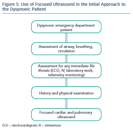
The authors recommend performing focused ultrasound during the initial encounter with the patient. Assuming critical life-saving interventions or immediate resuscitation are not required, focused ultrasound occurs immediately after the history, physical examination and ECG. As discussed in detail below, multiple studies demonstrate the utility of ultrasound to improve diagnostic accuracy in dyspnoeic ED patients.

Discussion Lung or Pulmonary Ultrasound: B-lines

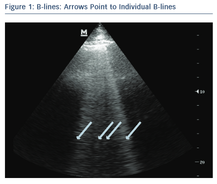
One of the most valuable and easiest to perform ultrasound assessments is lung ultrasound (LUS) looking for B-lines. LUS is performed using a low-frequency transducer – phased-array, microconvex or curvilinear probe – to allow for greater penetration and visualisation of B-lines.14,15 B-lines are vertical echogenic artifacts that originate from the pleural line, extend to the bottom of the ultrasound screen and move with lung sliding (see Figure 1).15,16 They are generated by water-thickened interlobular septa and may be seen in a variety of lung pathologies including pulmonary oedema.16–18 In the clinical setting of suspected AHF, pulmonary oedema is determined sonographically, using an eight-zone scanning technique, as greater than three B-lines in a rib space in at least two lung zones bilaterally.19,20 This finding on LUS is highly sensitive for the diagnosis of AHF, can be used to differentiate AHF from chronic obstructive pulmonary disease (COPD)14,16,18,21–24,10,25,26 and has better diagnostic accuracy for AHF than chest X-ray.15,27,28 In small studies, B-lines correlate well with natriuretic peptide levels8 and wedge pressure,28 though a more recent study shows only a modest correlation with filling pressures.29 In addition, LUS evaluation of B-lines is non-invasive, facile, feasible and has high inter-rater agreement.20,25 An alternative scanning protocol, counting the number of B-lines in 28 total rib spaces bilaterally, may allow for a more precise quantification of pulmonary oedema.30,31 In AHF patients, B-line assessment aids in diagnosis and may guide acute management.13,17 Protocols that combine lung and cardiac ultrasound have been found to be highly accurate for diagnosing AHF13,32 and potentially more accurate than LUS alone.13

Point to Individual B-Lines

Article image

Pleural Effusion : Seen Using Liver as Acoustic Window

Article image

Distended Inferior Vena Cava In Long Axis View

Article image

Pleural effusions

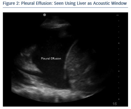
Pleural effusions are visualised sonographically as an anechoic or black fluid collection between the visceral and parietal pleura, in a dependent pattern (see Figure 2).15 Pleural effusions may be seen in AHF, as well as a variety of pulmonary pathologies including pneumonia and cancer.33 Literature is conflicting on whether the presence of a pleural effusion on LUS adds to the diagnosis of AHF. One study found pleural effusions were an unreliable finding in patients with AHF and less accurate than B-lines alone,25 while another study determined the presence of a pleural effusion combined with an ejection fraction <45 % to be highly specific for AHF and more accurate than B-line assessment alone.13

Cardiovascular Ultrasound: Inferior Vena Cava

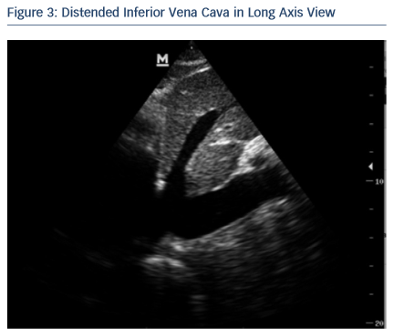
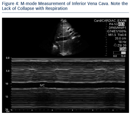
Measurement of inferior vena cava (IVC) diameter, along with its variability with respiration to determine intravascular volume status, has been described for decades.34 IVC measurement and changes with respiration correlate with right atrial and central venous pressures.35–38 As use of has become more widespread, measurement of IVC to determine fluid responsiveness in multiple patient populations has become more commonplace.39,40 IVC size and collapsibility index should be measured in long axis caudal to the hepatic vein inlet, or in short axis at the level of the left renal vein (see Figure 3).41 In general, IVC size and collapsibility with respiration is most valuable at the extremes (volume overloaded – no collapse during inspiration, or volume depleted – positive inspiratory collapse) as a qualitative finding (see Figure 4). In a euvolemic patient, the IVC partially collapses during inspiration, reflecting negative intrathoracic pressure. Importantly, this applies only to patients breathing spontaneously. In mechanically ventilated patients, the opposite occurs during a respiratory cycle; during expiration, the negative intrathoracic pressure leads to partial collapse in a euvolemic patient. In patients who present with acute dyspnoea, a minimal change in IVC diameter with respiration may predict volume overload and helps to differentiate AHF from COPD.42,43

Ejection Fraction (EF)

Ultrasound-trained emergency physicians can accurately determine left ventricular ejection fraction (LVEF) using both visual estimation and mitral E-point septal separation (EPSS) with good inter-rater reliability.44–46 Even with limited training, emergency physicians accurately estimate LVEF.47 However, EPSS is limited by septal wall motion abnormalities, mitral valve stenosis and angle of evaluation.48 Thus, the authors recommend EPSS be used in conjunction with other methods. If only a qualitative assessment of EF is desired, EPSS may be used to confirm visual estimation. International evidence-based recommendations advocate focused cardiac ultrasound be used to assess left ventricle dimensions and systolic function.6 Although a reduced EF in the setting of AHF signs and symptoms facilitates the diagnosis, the absence of a reduced EF does not rule it out. As nearly 50 % of patients with heart failure have a preserved EF, knowledge of the EF itself does not significantly alter acute management in the ER setting. The vast majority of AHF patients receive intravenous loop diuretics with a much smaller proportion receiving vasodilators. These treatments are given to AHF patients irrespective of EF and, at the present time, no robust evidence exists to suggest initial pharmacological management in the ER should differ based on EF.

Thus, ultrasound imaging of the heart may be more valuable to ‘rule-out’ other diagnoses rather than ‘ruling-in’ AHF. For example, presence of a significant pericardial effusion or right ventricular (RV) dilatation might suggest alternative diagnoses such as tamponade or pulmonary embolism.

Pitfalls

Ultrasound findings must be interpreted in clinical context. Although B-lines in the setting of a history and examination consistent with AHF most likely represent pulmonary congestion, B-lines lack specificity and are seen in multiple other conditions; for example, non-cardiogenic pulmonary oedema, pulmonary fibrosis, pneumonia, pneumonitis, and lung cancer.18,22,25 Ensuring that B-lines are bilateral helps to differentiate AHF from other etiologies. Absence of B-lines in one hemi-thorax greatly lowers the probability of AHF.

Some limitations in determining volume overload by measuring IVC diameter variability include etiologies that increase right atrial pressure outside of acute heart failure, such as pulmonary emboli, pulmonary hypertension, and cardiac tamponade. These examples highlight the importance of clinical context, as well as radiographic and biomarker features. In addition, these examples reveal the potential pitfall of examining the IVC only, rather than examining additional views of the lung and the heart.

As nearly half of heart failure patients have a relatively preserved EF, the condition is not excluded despite a normal EF.

While LUS is one of the easiest assessments to perform, IVC and LVEF evaluation may require some additional training; however, this is still well within the realm of many specialties.49,50 Importantly, focused cardiac ultrasound does not replace formal ECG;5 instead, focused bedside ultrasound aids in the diagnosis and management of many conditions, including AHF.

M-mode Measurement of Inferior Vena Cava

Article image

Focused Ultrasound in the Initial Approach

Article image

Future Directions

Multiple studies support the added value of a focused ultrasound to facilitate a diagnosis of AHF, but it remains infrequently used. As ultrasound devices become increasingly smaller and familiarity with focused ultrasound increases, its use will continue to grow. However, there are limited studies evaluating the utility of ultrasound to guide management and prognosis in the ER setting. Small studies have shown the efficacy of LUS to measure resolution of extra-vascular lung water or B-lines.17,51 Ongoing work in this area may demonstrate a future role for ultrasound to guide the management of AHF.

Conclusions

The ED plays a pivotal role in the initial management of AHF. Correct diagnosis and treatment is critical to ensure the right tone is set for subsequent in-hospital or outpatient management. As patients present with symptoms and signs and not diagnoses, AHF may be missed, especially in patients with other co-morbid conditions. Focused ultrasound is a fast, non-invasive method that rapidly aids the clinician to rule in or rule out various cardiac and pulmonary diagnoses. Importantly, focused ultrasound must be interpreted within the clinical context. For potential AHF patients, the use of LUS to assess for B-lines and pleural effusions, cardiac ultrasound to assess EF and measure IVC diameter and respiratory variation are skills that may be learned quickly and can aid the clinician.

Clinical Case Study Conclusions

Given the patient’s history of coronary artery disease, AHF is on the differential diagnosis list – but so are acute coronary syndrome, COPD, and pneumonia. The ECG shows sinus tachycardia, but no other acute ischaemic changes. While awaiting chest X-ray, a focused ultrasound is performed, starting with the lungs (see Figure 5). Four or five B-lines are noted in at least two zones bilaterally, raising suspicion of pulmonary congestion. There are no pleural effusions. Cardiac examination does not show any pericardial effusion or RV dilation and systolic function appears mildly depressed by qualitative assessment. IVC assessment is difficult given the patient’s habitus, but minimal respiratory collapse is observed, suggesting elevated venous pressures. Based on this evidence, treatment is given for AHF. Chest X-ray reveals mild cardiomegaly, mild vascular engorgement, no Kerley B-lines, and no consolidation.

References

  1. Gheorghiade M, Pang PS. Acute heart failure syndromes. J Am Coll Cardiol 2009;53:557–73.
    Crossref | PubMed
  2. Mebazaa A, Pang PS, Tavares M, et al. The impact of early standard therapy on dyspnoea in patients with acute heart failure: the URGENT-dyspnoea study. Eur Heart J 2010;31:832–41.
    Crossref | PubMed
  3. ADHERE Scientific Advisory Committee. Acute Decompensated Heart Failure National Registry(ADHERE®) Core Module Q1 2006 Final Cumulative National Benchmark Report. Scios, Inc; July 2006.
  4. Labovitz AJ, Noble VE, Bierig M, et al. Focused cardiac ultrasound in the emergent setting: a consensus statement of the American Society of Echocardiography and American College of Emergency Physicians. J Am Soc Echocardiogr 2010;23:1225–30.
    Crossref | PubMed
  5. Neskovic AN, Edvardsen T, Galderisi M, et al. Focus cardiac ultrasound: the European Association of Cardiovascular Imaging viewpoint. Eur Heart J Cardiovasc Imaging 2014;15:956–60.
    Crossref | PubMed
  6. Via G, Hussain A, Wells M, et al. International evidence-based recommendations for focused cardiac ultrasound. J Am Soc Echocardiogr 2014;27:683.e1–683 e33.
    Crossref | PubMed
  7. Fields M. AEUS Narrated Lecture Series. AEUS Narrated Lecture Series. 2014. Available at http://community.saem.org/blogs/matt-fields/2014/09/07/aeus-narrated-lecture-series (accessed 15 July 2015).
  8. Liteplo AS, Marill KA, Villen T, et al. Emergency thoracic ultrasound in the differentiation of the etiology of shortness of breath (ETUDES): sonographic B-lines and N-terminal pro- brain-type natriuretic peptide in diagnosing congestive heart failure. Acad Emerg Med 2009;16:201–10.
    Crossref | PubMed
  9. Martindale JL, Noble VE, Liteplo A. Diagnosing pulmonary edema: lung ultrasound versus chest radiography. Eur J Emerg Med 2013;20:356–360.
    Crossref | PubMed
  10. Gargani L, Frassi F, Soldati G, et al. Ultrasound lung comets for the differential diagnosis of acute cardiogenic dyspnoea: a comparison with natriuretic peptides. Eur J Heart Fail 2008;10:70–7.
    Crossref | PubMed
  11. Picano E, Gargani L, Gheorghiade M. Why, when, and how to assess pulmonary congestion in heart failure: pathophysiological, clinical, and methodological implications. Heart Fail Rev 2010;15:63–72.
    Crossref | PubMed
  12. Soldati G, Gargani L, Silva FR. Acute heart failure: new diagnostic perspectives for the emergency physician. Intern Emerg Med 2008;3:37–41.
    Crossref | PubMed
  13. Russell FM, Ehrman RR, Cosby K, et al. Diagnosing acute heart failure in patients with undifferentiated dyspnea: a lung and cardiac ultrasound (LuCUS) protocol. Acad Emerg Med 2015;22:182–91.
    Crossref | PubMed
  14. Chiem AT, Chan CH, Ander DS, et al. Comparison of expert and novice sonographers’ performance in Focused Lung Ultrasonography in Dyspnea (FLUID) to diagnose patients with acute heart failure syndrome. Acad Emerg Med 2015;22:564–73.
    Crossref | PubMed
  15. Volpicelli G, Elbarbary M, Blaivas M, et al. International evidence-based recommendations for point-of-care lung ultrasound. Intensive Care Med 2012;38:577–91.
    Crossref | PubMed
  16. Lichtenstein DA, Meziere GA. Relevance of lung ultrasound in the diagnosis of acute respiratory failure: the BLUE protocol. Chest 2008;134:117–25.
    Crossref | PubMed
  17. Volpicelli G, Caramello V, Cardinale L, et al. Bedside ultrasound of the lung for the monitoring of acute decompensated heart failure. Am J Emerg Med 2008;26: 585–91.
    Crossref | PubMed
  18. Volpicelli G, Cardinale L, Garofalo G, Veltri A. Usefulness of lung ultrasound in the bedside distinction between pulmonary edema and exacerbation of COPD. Emerg Radiol 2008;15:145–51.
    Crossref | PubMed
  19. Volpicelli G, Silva F, Radeos M. Real-time lung ultrasound for the diagnosis of alveolar consolidation and interstitial syndrome in the emergency department. Eur J Emerg Med 2010;17:63–72.
    Crossref | PubMed
  20. Noble VE, Lamhaut L, Capp R, et al. Evaluation of a thoracic ultrasound training module for the detection of pneumothorax and pulmonary edema by prehospital physician care providers. BMC Med Educ 2009;9:3.
    Crossref | PubMed
  21. Al Deeb M, Barbic S, Featherstone R, et al. Point-of-care ultrasonography for the diagnosis of acute cardiogenic pulmonary edema in patients presenting with acute dyspnea: a systematic review and meta-analysis. Acad Emerg Med 2014;21:843–52.
    Crossref | PubMed
  22. Lichtenstein D, Meziere G. A lung ultrasound sign allowing bedside distinction between pulmonary edema and COPD: the comet-tail artifact. Intensive Care Med 1998;24:1331–4.
    Crossref | PubMed
  23. Lichtenstein D, Meziere G, Biderman P, et al. The comet-tail artifact. An ultrasound sign of alveolar-interstitial syndrome. Am J Respir Crit Care Med 1997;156:1640–6.
    Crossref | PubMed
  24. Lichtenstein DA. Ultrasound in the management of thoracic disease. Crit Care Med 2007;35:S250–61.
    Crossref | PubMed
  25. Cibinel GA, Casoli G, Elia F, et al. Diagnostic accuracy and reproducibility of pleural and lung ultrasound in discriminating cardiogenic causes of acute dyspnea in the emergency department. Intern Emerg Med 2012;7:65–70.
    Crossref | PubMed
  26. Pivetta E, Goffi A, Lupia E, et al. Lung ultrasound- implemented diagnosis of acute decompensated heart failure in the ED: A SIMEU Multicenter Study. Chest 2015;148:202–10.
    Crossref | PubMed
  27. Xirouchaki N, Magkanas E, Vaporidi K, et al. Lung ultrasound in critically ill patients: comparison with bedside chest radiography. Intensive Care Med 2011;37:1488–93.
    Crossref | PubMed
  28. Agricola E, Bove T, Oppizzi M, et al. Ultrasound comet-tail images: a marker of pulmonary edema: a comparative study with wedge pressure and extravascular lung water. Chest 2005;127:1690–5.
    Crossref | PubMed
  29. Platz E, Lattanzi A, Agbo C, et al. Utility of lung ultrasound in predicting pulmonary and cardiac pressures. Eur J Heart Fail 2012;14:1276–84.
    Crossref | PubMed
  30. Jambrik Z, Monti S, Coppola V, et al. Usefulness of ultrasound lung comets as a nonradiologic sign of extravascular lung water. Am J Cardiol 2004;93:1265–70.
    Crossref | PubMed
  31. Picano E, Frassi F, Agricola E, et al. Ultrasound lung comets: a clinically useful sign of extravascular lung water. J Am Soc Echocardiogr 2006;19:356–63.
    Crossref | PubMed
  32. Kajimoto K, Madeen K, Nakayama T, et al. Rapid evaluation by lung-cardiac-inferior vena cava (LCI) integrated ultrasound for differentiating heart failure from pulmonary disease as the cause of acute dyspnea in the emergency setting. Cardiovasc Ultrasound 2012;10:49.
    Crossref | PubMed
  33. Woodring JH. Distribution of pleural effusion in congestive heart failure: what is atypical? South Med J 2005;98:518–23.
    Crossref | PubMed
  34. Natori H, Tamaki S, Kira S. Ultrasonographic evaluation of ventilatory effect on inferior vena caval configuration. Am Rev Respir Dis 1979;120:421–7.
    PubMed
  35. Moreno FL, Hagan AD, Holmen JR, et al. Evaluation of size and dynamics of the inferior vena cava as an index of right- sided cardiac function. Am J Cardiol 1984;53:579–85.
    Crossref | PubMed
  36. Kircher BJ, Himelman RB, Schiller NB. Noninvasive estimation of right atrial pressure from the inspiratory collapse of the inferior vena cava. Am J Cardiol 1990;66:493–6.
    Crossref | PubMed
  37. Bodson L, Vieillard-Baron A. Respiratory variation in inferior vena cava diameter: surrogate of central venous pressure or parameter of fluid responsiveness? Let the physiology reply. Crit Care 2012;16:181.
    Crossref | PubMed
  38. Citilcioglu S, Sebe A, Ay MO, et al. The relationship between inferior vena cava diameter measured by bedside ultrasonography and central venous pressure value. Pak J Med Sci 2014;30:310–5.
    Crossref | PubMed
  39. Muller L, Bobbia X, Toumi M, et al. Respiratory variations of inferior vena cava diameter to predict fluid responsiveness in spontaneously breathing patients with acute circulatory failure: need for a cautious use. Crit Care 2012;16:R188.
    Crossref | PubMed
  40. Moretti R, Pizzi B. Inferior vena cava distensibility as a predictor of fluid responsiveness in patients with subarachnoid hemorrhage. Neurocrit Care 2010;13:3–9.
    Crossref | PubMed
  41. Wallace DJ, Allison M, Stone MB. Inferior vena cava percentage collapse during respiration is affected by the sampling location: an ultrasound study in healthy volunteers. Acad Emerg Med 2010;17:96–9.
    Crossref | PubMed
  42. Anderson KL, Jenq KY, Fields JM, et al. Point-of-care ultrasound diagnoses acute decompensated heart failure in the ED regardless of examination findings. Am J Emerg Med 2014;32:385–8.
    Crossref | PubMed
  43. Blehar DJ, Dickman E, Gaspari R. Identification of congestive heart failure via respiratory variation of inferior vena cava diameter. Am J Emerg Med 2009;27:71–5.
    Crossref | PubMed
  44. Moore CL, Rose GA, Tayal VS, et al. Determination of left ventricular function by emergency physician echocardiography of hypotensive patients. Acad Emerg Med 2002;9:186–93.
    Crossref | PubMed
  45. Randazzo MR, Snoey ER, Levitt MA, Binder K. Accuracy of emergency physician assessment of left ventricular ejection fraction and central venous pressure using echocardiography. Acad Emerg Med 2003;10:973–7.
    Crossref | PubMed
  46. Secko MA, Lazar JM, Salciccioli LA, Stone MB. Can junior emergency physicians use E-point septal separation to accurately estimate left ventricular function in acutely dyspneic patients? Acad Emerg Med 2011;18:1223–6.
    Crossref | PubMed
  47. Bustam A, Noor Azhar M, Singh Veriah R, et al. Performance of emergency physicians in point-of-care echocardiography following limited training. Emerg Med J 2014;31:369–73.
    Crossref | PubMed
  48. McKaigney CJ, Krantz MJ, La Rocque CL, et al. E-point septal separation: a bedside tool for emergency physician assessment of left ventricular ejection fraction. Am J Emerg Med 2014;32:493–7.
    Crossref | PubMed
  49. Unluer EE, Karagoz A, Oyar O, et al. Lung ultrasound by emergency nursing as an aid for rapid triage of dyspneic patients: a pilot study. Int Emerg Nurs 2014;22:226–31.
    Crossref | PubMed
  50. Kennedy Hall M, Coffey EC, Herbst M, et al. The “5Es” of emergency physician-performed focused cardiac ultrasound: a protocol for rapid identification of effusion, ejection, equality, exit, and entrance. Acad Emerg Med 2015;22:583–93.
    Crossref | PubMed
  51. Noble VE, Murray AF, Capp R, DJ, et al. Ultrasound assessment for extravascular lung water in patients undergoing hemodialysis. Time course for resolution. Chest 2009;135:1433–9.
    Crossref | PubMed