Article

Link Between Synovial and Myocardial Inflammation: Conceptual Framework to Explain the Pathogenesis of Heart Failure with Preserved Ejection Fraction in Patients with Systemic Rheumatic Diseases

Register or Login to View PDF Permissions
Permissions× For commercial reprint enquiries please contact Springer Healthcare: ReprintsWarehouse@springernature.com.

For permissions and non-commercial reprint enquiries, please visit Copyright.com to start a request.

For author reprints, please email rob.barclay@radcliffe-group.com.
Average (ratings)
No ratings
Your rating

Abstract

Patients with a broad range of systemic rheumatic diseases are at increased risk of heart failure (HF), an event that is not related to traditional cardiovascular risk factors or underlying ischaemic heart disease. The magnitude of risk is linked to the severity of arthritic activity, and HF is typically accompanied by a preserved ejection fraction. Subclinical evidence for myocardial fibrosis, microcirculatory dysfunction and elevated cardiac filling pressures is present in a large proportion of patients with rheumatic diseases, particularly those with meaningful systemic inflammation. Drugs that act to attenuate pro-inflammatory pathways (methotrexate and antagonists of tumour necrosis factor and interleukin-1) may ameliorate myocardial inflammation and cardiac structural abnormalities and reduce the risk of HF events.

Disclosure:The author has no conflicts of interest to declare

Received:

Accepted:

Correspondence Details:Milton Packer, Baylor Heart and Vascular Institute, Baylor University Medical Center, 621 North Hall St, Dallas, TX 75226, US. E: milton.packer@baylorhealth.edu

Copyright Statement:

The copyright in this work belongs to Radcliffe Medical Media. Only articles clearly marked with the CC BY-NC logo are published with the Creative Commons by Attribution Licence. The CC BY-NC option was not available for Radcliffe journals before 1 January 2019. Articles marked ‘Open Access’ but not marked ‘CC BY-NC’ are made freely accessible at the time of publication but are subject to standard copyright law regarding reproduction and distribution. Permission is required for reuse of this content.

Patients with a broad range of systemic inflammatory rheumatic diseases (i.e. rheumatoid arthritis, systemic lupus erythematosus, psoriatic arthritis and ankylosing spondylitis) are exceptionally prone to cardiovascular disorders, but previous work concerning the development of heart disease in these individuals has largely focused on the risk of MI.

However, the most important cardiovascular complication in these individuals is the development of heart failure (HF), an event that may not be readily apparent to many clinicians and is not related to traditional cardiovascular risk factors or to clinically evident ischaemic heart disease.1,2 HF develops with increasing rapidity following a diagnosis of systemic rheumatic diseases, and the magnitude of risk is related to the severity of arthritic activity.2 Typically, the four principal systemic rheumatic disorders, that is, rheumatoid arthritis, systemic lupus erythematosus, psoriatic arthritis and ankylosing spondylitis, increase the risk of new-onset HF as much as two- to threefold, when compared with the general population.2–9

Importantly, both the clinical presentation and the outcome of HF differ between patients with and without these systemic rheumatic diseases. Although patients with systemic rheumatic diseases are prone to MI (a form of injury that is typically linked to HF with reduced ejection fraction [HFrEF]), the phenotype of HF in most patients with systemic rheumatic diseases is characterised by the absence of coronary artery disease (CAD) and a preserved EF, with its characteristic features of diastolic filling abnormalities, microvascular dysfunction and cardiac fibrosis on cardiac imaging.10–17 This finding is not surprising; of the two main phenotypes of HF, that is, HFrEF or HF with preserved EF (HFpEF), the former is related to cardiomyocyte loss and stretch, whereas the latter is primarily related to the presence of systemic inflammatory disorders, most commonly, obesity and diabetes.18–20

Mechanisms by Which Systemic Inflammatory Disorders Cause the Phenotype of HFpEF

Systemic inflammatory disorders can target the microcirculation of the myocardium, leading to microvascular endothelial dysfunction and fibrosis. The inflammatory injury can directly affect the coronary microvasculature; additionally, its effect can be mediated indirectly, because systemic inflammation promotes the expansion and biological transformation of epicardial adipose tissue (EAT), which acts as an amplifier to focus the systemic inflammatory process onto the underlying myocardium, with which it shares an unobstructed microcirculation.21,22 In healthy people, epicardial fat has the biological properties of brown adipose tissue, which combusts pro-inflammatory fatty acids and secretes adipokines (e.g. adiponectin) that suppress inflammation and nourish the heart. However, in the presence of systemic inflammation, mesenchymal cells in the epicardium proliferate and develop features of white adipose tissue, which is prone to lipolysis and causes the release of fatty acids that trigger infiltration of the tissues by macrophages, as well as the secretion of pro-inflammatory cytokines (e.g. leptin, tumour necrosis factor [TNF]-alpha, interleukin [IL]-6, IL-1beta and resistin).22–25

Postulated Mechanisms

Article image

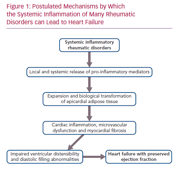
It is therefore noteworthy that EAT mass is increased in patients with rheumatoid arthritis, systemic lupus erythematosus, psoriatic arthritis and ankylosing spondylitis.26–30 The intimacy of its interface with the myocardium allows the biological derangements in epicardial fat to be readily transmitted to the neighbouring muscle.23 Acting locally, pro-inflammatory adipocytokines synthesised in and released from epicardial fat depots can cause a spread of the inflammatory process to underlying tissues, which includes the coronary arteries, as well as the atrial and ventricular myocardium, leading to the epicardial adipose inflammatory triad: coronary atherosclerosis, AF and HFpEF.31 The extension of inflammation to the perivascular tissues surrounding the coronary arteries likely explains the increased risk of MI in rheumatoid arthritis, systemic lupus erythematosus, psoriatic arthritis and ankylosing spondylitis.32–34 The spread of inflammation to myocardial tissues leads to microcirculatory dysfunction and rarefaction, as well as to increased deposition of collagen.35,36 MRI in patients with heightened quantities of epicardial fat reveals increases in extracellular volume, indicative of underlying myocardial fibrosis.36 If the process abuts the atria, the resulting electroanatomical remodelling and fragmentation leads to AF, thus explaining the increased incidence and prevalence of the arrhythmia in rheumatoid arthritis, systemic lupus erythematosus, psoriatic arthritis and ankylosing spondylitis.37–40 Finally, if the abnormalities in EAT adjoin the left ventricle, the microvascular dysfunction and cardiac fibrosis act together to impair the distensibility of the chamber, thereby limiting its ability to accommodate blood volume, but without a decrease in systolic function as measured by EF.41,42 The result is the HF phenotype known as HFpEF (Figure 1).

Mediating Effects of Leptin, Aldosterone and Natriuretic Peptides in the Pro-inflammatory Expansion of EAT

Why does systemic inflammation cause an expansion and pro-inflammatory transformation of EAT? The interactions among several endogenous hormonal mediators that are known to play a role in systemic inflammation (i.e. aldosterone, natriuretic peptides and leptin) contribute importantly to the development of epicardial adiposity and inflammation.4,5 Aldosterone promotes adipogenesis and causes an expansion and pro-inflammatory transformation of EAT,43,44 whereas epicardial adiposity is inversely related to the level of endogenous natriuretic peptides,45 presumably because natriuretic peptide signalling antagonises adipocyte hypertrophy.46 Additionally, aldosterone promotes pro-inflammatory pathways;47 mineralocorticoid receptor antagonism attenuates inflammasome activity and blocks the production of pro-inflammatory cytokines in adipocytes and macrophages.48 Conversely, endogenous natriuretic peptides also play an important role in the pathogenesis of systemic inflammatory disorders, but in a manner that opposes the actions of aldosterone. Natriuretic peptides inhibit biological pathways involved in inflammation and attenuate the production of pro-inflammatory cytokines in macrophages and adipocytes.49,50 Finally, EAT secretes leptin, which is also known to play a central role in immune responses and inflammation.51 Leptin mediates the proliferation of monocytes and their production of pro-inflammatory cytokines;51,52 circulating levels of the adipokine are elevated in systemic inflammatory states and are directly related to epicardial fat volume.53,54

Patients with obesity have increased circulating levels of aldosterone and leptin and decreased levels of natriuretic peptides.55–57 These relationships can be explained by the secretion of both aldosterone and leptin by an expanded mass of adipocytes,58 as well as by an ability of adipocytes to enhance the degradation of natriuretic peptides, either through an action to secrete neprilysin or to enhance peptide clearance.59,60 The increase in aldosterone and leptin (accompanied by the decrease in natriuretic peptides) acts to promote the systemic pro-inflammatory state. The interplay of these mediators may explain why obesity increases the incidence, enhances the clinical and radiographic severity and attenuates the degree of symptomatic improvement following the use of anti-inflammatory treatment in patients with rheumatoid arthritis, systemic lupus erythematosus, psoriatic arthritis and ankylosing spondylitis.61–68

In patients with expanded and dysfunctional EAT, the release of leptin and aldosterone can promote inflammation, microcirculatory dysfunction and fibrosis in the underlying myocardium,69 effects that are opposed by the anti-inflammatory and antifibrotic actions of endogenous natriuretic peptides.70 The result of these interactions is an impairment of the distensibility of the left ventricle, such that blood volume is accommodated only at the expense of a disproportionate increase in cardiac filling pressures, thus leading to exertional dyspnoea and HF. These observations explain why patients with HFpEF have elevated circulating levels of leptin and aldosterone but inappropriately suppressed levels of natriuretic peptides.25,42,71 Interference with the actions of aldosterone (and possibly leptin), as well as potentiation of natriuretic peptides, has yielded structural and clinical benefits in patients with HFpEF in randomised controlled trials.72–77

Activation of Pathogenetic Mechanisms for HFpEF in Systemic Rheumatic Diseases

Both rheumatoid arthritis and ankylosing spondylitis are characterised by increased levels of aldosterone in circulating blood and inflamed tissues,78,79 and spironolactone’s anti-inflammatory actions have been proposed as a treatment for these arthritides, as well as for lupus nephritis.80,81 At the same time, levels of leptin in blood (and often synovial fluid) are increased in rheumatoid arthritis, systemic lupus erythematosus, psoriatic arthritis and ankylosing spondylitis; are associated with the degree of joint and skeletal involvement; and are linked with patient-reported outcomes and measures of disease activity.82–93 Interestingly, high levels of leptin may identify patients with rheumatic diseases who respond poorly to anti-inflammatory treatment.94 Interestingly, increased leptin signalling may play a direct pathogenetic role in both rheumatoid arthritis and systemic lupus erythematosus by increasing the production of pro-inflammatory cytokines and autoantibodies and inhibiting immune regulation;95–99 interference with leptin signalling ameliorates the course of experimental lupus.100

Finally, the activity of neprilysin is increased at sites of joint involvement in rheumatoid arthritis,101 where it may play a role in degrading locally active natriuretic peptides and limiting their counterbalancing anti-inflammatory and anti-adipogenic actions. The release of neprilysin from the expanded mass of epicardial adipocytes may further diminish the ability of natriuretic peptides to minimise local cardiac fibrosis.102 This accelerated degradation of biologically active natriuretic peptides should not be confused with increased circulating levels of N-terminal pro-B-type natriuretic peptide (an inactive prohormone) in systemic rheumatic diseases; the prohormone is indicative of cardiac stress and is not degraded by neprilysin. As a result of these proadipogenic and pro-inflammatory interactions, EAT is increased in patients with rheumatoid arthritis, systemic lupus erythematosus, psoriatic arthritis and ankylosing spondylitis, and the magnitude of increase is generally proportional to the duration and severity of the underlying systemic inflammatory process.26–30

The transmission of epicardial inflammation to the underlying myocardial tissues explains why many patients with systemic rheumatic disease have coronary microcirculatory dysfunction, cardiac fibrosis, ventricular diastolic filling abnormalities, left atrial enlargement and HF in the absence of any evidence for or a history of MI or myocardial injury.1,11–17,26,103–110 Subclinical evidence for myocardial fibrosis on cardiac MRI and echocardiographic features of elevated cardiac filling pressures (as reflected by abnormal diastolic filling dynamics or abnormal left atrial geometry, and derangements in coronary microcirculatory dysfunction) are present in 30–50% of patients with rheumatoid arthritis, systemic lupus erythematosus, psoriatic arthritis and ankylosing spondylitis, particularly those with meaningful systemic inflammatory activity, and these cardiac structural and functional abnormalities parallel the severity and duration of joint involvement.26,105–107,110–112 These are the hallmarks of the ventricular myopathy that underlies the development of HFpEF, and they are often undiagnosed in clinical practice.

Effect of Anti-cytokine Agents and Neurohormonal Antagonists on the Development of HF In Systemic Rheumatic Disorders

If the proposed framework is valid, then interventions that directly interrupt inflammatory pathways might be useful in preventing and treating HFpEF in patients with systemic rheumatic disorders. Methotrexate exerts anti-inflammatory effects in adipose tissue and reduces cardiac fibrosis following experimental cardiac injury.113 Its use in rheumatic disorders reduces pro-inflammatory cytokines and has been associated with a lower risk of HF hospitalisations.114

The IL-1 family has been implicated in adipose tissue inflammation, and experimental inhibition has been accompanied by decreases in pro-inflammatory cytokines and cardiac fibrosis. In a small cross-over trial, the IL-1 antagonist anakinra reduced systemic inflammation and improved exercise capacity in patients with HFpEF.115 More impressively, in a randomised controlled trial of >10,000 patients with evidence for systemic inflammation, long-term treatment with the IL-1beta antagonist canakinumab decreased inflammatory biomarkers and the risk of hospitalisation for HF.116

The use of antagonists of TNF-alpha in the management of patients with rheumatic diseases (who are prone to HFpEF) has been associated with amelioration of myocardial inflammation, coronary microvascular dysfunction and cardiac functional abnormalities, and little change in or a reduced risk of HF events.117–121 These findings stand in contrast to concerns that the use of antagonists of TNF-alpha may exacerbate the clinical course of HFrEF (as is commonly seen following MI).122,123

If activation of the leptin-aldosterone-neprilysin axis plays a role in mediating the pro-inflammatory expansion of EAT,25 then drugs that modulate this axis could influence the course of HFpEF in patients who have rheumatoid arthritis, systemic lupus erythematosus, psoriatic arthritis and ankylosing spondylitis. Trials of mineralocorticoid receptor antagonists and neprilysin inhibitors in patients with HFpEF have not specifically targeted patients with systemic rheumatic disorders. However, it is noteworthy that these systemic rheumatic disorders are particularly common in women;124–127 and on subgroup analysis the benefits of mineralocorticoid receptor antagonists and neprilysin inhibitors have been reported to be greater in women than in men.74,128

Conclusion

Many systemic inflammatory rheumatic diseases, that is, rheumatoid arthritis, systemic lupus erythematosus, psoriatic arthritis and ankylosing spondylitis, are accompanied by an increased risk of HF, which begins at the onset of diagnosis and increases in proportion to the clinical severity and duration of the systemic inflammatory process. HF is not the result of accelerated CAD and ischaemic myocardial injury, but instead is related to myocardial inflammation, coronary microvascular dysfunction and fibrosis leading to HFpEF. A conceptual framework is proposed to explain the evolution of HF in these patients. Pro-inflammatory mediators characteristic of systemic inflammatory states (e.g. leptin, aldosterone and neprilysin) can cause expansion and biological transformation of EAT, leading to coronary microvascular injury and fibrosis of the underlying myocardium. EAT volume is increased in these systemic rheumatic disorders, and 30–50% of afflicted patients have subclinical evidence of cardiac inflammation, microcirculatory dysfunction and fibrosis, as well as the structural and functional features of HFpEF. Current anti-inflammatory agents that are used for the treatment of these systemic rheumatic diseases have the potential to minimise the development of cardiac involvement and thereby reduce the risk of HF. In addition, it is possible that drugs that modulate the leptin-aldosterone-neprilysin axis could modify the cardiovascular consequences of systemic inflammation and change the clinical course of HFpEF in these patients.

References

  1. Nicola PJ, Maradit-Kremers H, Roger VL, et al. The risk of congestive heart failure in rheumatoid arthritis: a population-based study over 46 years. Arthritis Rheum 2005;52:412–20.
    Crossref | PubMed
  2. Mantel Ä, Holmqvist M, Andersson DC, et al. Association between rheumatoid arthritis and risk of ischemic and nonischemic heart failure. J Am Coll Cardiol 2017;69:1275–85.
    Crossref | PubMed
  3. Zou K, Xiao FK, Li HY, et al. Risk of cardiovascular disease in Chinese patients with rheumatoid arthritis: a cross-sectional study based on hospital medical records in 10 years. PLoS One 2017;12:e0180376.
    Crossref | PubMed
  4. Kim CH, Al-Kindi SG, Jandali B, et al. Incidence and risk of heart failure in systemic lupus erythematosus. Heart 2017;103:227–33.
    Crossref | PubMed
  5. Kibari A, Cohen AD, Gazitt T, et al. Cardiac and cardiovascular morbidities in patients with psoriatic arthritis: a population-based case control study. Clin Rheumatol 2019;38:2069–75.
    Crossref | PubMed
  6. Polachek A, Touma Z, Anderson M, Eder L. Risk of cardiovascular morbidity in patients with psoriatic arthritis: a meta-analysis of observational studies. Arthritis Care Res (Hoboken) 2017;69:67–74.
    Crossref | PubMed
  7. Han C, Robinson DW Jr, Hackett MV, et al. Cardiovascular disease and risk factors in patients with rheumatoid arthritis, psoriatic arthritis, and ankylosing spondylitis. J Rheumatol 2006;33:2167–72.
    PubMed
  8. Szabo SM, Levy AR, Rao SR, et al. Increased risk of cardiovascular and cerebrovascular diseases in individuals with ankylosing spondylitis: a population-based study. Arthritis Rheum 2011;63:3294–304.
    Crossref | PubMed
  9. Bae KH, Hong JB, Choi YJ, et al. Association of congestive heart failure and death with ankylosing spondylitis: a nationwide longitudinal cohort study in Korea. J Korean Neurosurg Soc 2019;62:217–24.
    Crossref | PubMed
  10. Davis JM 3rd, Roger VL, Crowson CS, et al. The presentation and outcome of heart failure in patients with rheumatoid arthritis differs from that in the general population. Arthritis Rheum 2008;58:2603–11.
    Crossref | PubMed
  11. Amigues I, Russo C, Giles JT, et al. Myocardial microvascular dysfunction in rheumatoid arthritis. Quantitation by 13N-ammonia positron emission tomography/computed tomography. Circ Cardiovasc Imaging 2019;12:e007495.
    Crossref | PubMed
  12. Elnady BM, Abdelghafar AS, Khalik ES, et al. The implication of tissue Doppler echocardiography and cardiopulmonary exercise in early detection of cardiac dysfunction in systemic lupus erythematosus patients. Eur J Rheumatol 2016;3:109–17.
    Crossref | PubMed
  13. Heslinga SC, Van Dongen CJ, Konings TC, et al. Diastolic left ventricular dysfunction in ankylosing spondylitis: a systematic review and meta-analysis. Semin Arthritis Rheum 2014;44:14–9.
    Crossref | PubMed
  14. Sandhu VK, Wei J, Thomson LEJ, et al. A five-year follow up of coronary microvascular dysfunction and coronary artery disease in SLE: results from a community-based lupus cohort. Arthritis Care Res (Hoboken) 2019; epub ahead of press.
    Crossref | PubMed
  15. Tufano A, Lembo M, Di Minno MN, et al. Left ventricular diastolic abnormalities other than valvular heart disease in antiphospholipid syndrome: an echocardiographic study. Int J Cardiol 2018;271:366–70.
    Crossref | PubMed
  16. Shang Q, Tam LS, Sanderson JE, et al. Impaired left ventricular apical rotation is associated with disease activity of psoriatic arthritis. J Rheumatol 2014;41:706–13.
    Crossref | PubMed
  17. Shang Q, Tam LS, Yip GW, et al. High prevalence of subclinical left ventricular dysfunction in patients with psoriatic arthritis. J Rheumatol 2011;38:1363–70.
    Crossref | PubMed
  18. Paulus WJ, Dal Canto E. Distinct myocardial targets for diabetes therapy in heart failure with preserved or reduced ejection fraction. JACC Heart Fail 2018;6:1–7.
    Crossref | PubMed
  19. Packer M. Critical role of the epicardium in mediating cardiac inflammation and fibrosis in patients with type 2 diabetes. Diabetes Obes Metab 2019;21:1765–8.
    Crossref | PubMed
  20. Packer M, Kitzman DW. Obesity-related heart failure with a preserved ejection fraction: the mechanistic rationale for combining inhibitors of aldosterone, neprilysin, and sodium-glucose cotransporter-2. JACC Heart Fail 2018;6:633–9.
    Crossref | PubMed
  21. Paulus WJ, Tschöpe C. A novel paradigm for heart failure with preserved ejection fraction: comorbidities drive myocardial dysfunction and remodeling through coronary microvascular endothelial inflammation. J Am Coll Cardiol 2013;62:263–71.
    Crossref | PubMed
  22. Packer M. Epicardial adipose tissue may mediate deleterious effects of obesity and inflammation on the myocardium. J Am Coll Cardiol 2018;71:2360–72.
    Crossref | PubMed
  23. Greulich S, Maxhera B, Vandenplas G, et al. Secretory products from epicardial adipose tissue of patients with type 2 diabetes mellitus induce cardiomyocyte dysfunction. Circulation 2012;126:2324–34.
    Crossref | PubMed
  24. Mazurek T, Zhang L, Zalewski A, et al. Human epicardial adipose tissue is a source of inflammatory mediators. Circulation 2003;108:2460–6.
    Crossref | PubMed
  25. Packer M. Leptin-aldosterone-neprilysin axis: identification of its distinctive role in the pathogenesis of the three phenotypes of heart failure in people with obesity. Circulation 2018;137:1614–31.
    Crossref | PubMed
  26. Alpaydın S, Buyukterzi Z, Akkurt HE, Yılmaz H. Impaired left ventricular diastolic functions and thickened epicardial adipose tissue in rheumatoid arthritis patients is correlated with DAS-28 score. Acta Cardiol Sin 2017;33:182–7.
    Crossref | PubMed
  27. Lipson A, Alexopoulos N, Hartlage GR, et al. Epicardial adipose tissue is increased in patients with systemic lupus erythematosus. Atherosclerosis 2012;223:389–93.
    Crossref | PubMed
  28. Wang X, Guo Z, Zhu Z, et al. Epicardial fat tissue in patients with psoriasis: a systematic review and meta-analysis. Lipids Health Dis 2016;15:103.
    Crossref | PubMed
  29. Çag˘lar SO, Boyraz I., Erdem F, et al. Evaluation of atrial conduction times, epicardial fat thickness and carotid intima-media thickness in patients with ankylosing spondylitis. Arch Rheumatol 2016;31:353–8.
    Crossref | PubMed
  30. Resorlu H, Akbal A, Resorlu M, et al. Epicardial adipose tissue thickness in patients with ankylosing spondylitis. Clin Rheumatol 2015;34:295–9.
    Crossref | PubMed
  31. Packer M. The epicardial adipose inflammatory triad: coronary atherosclerosis, atrial fibrillation, and heart failure with a preserved ejection fraction. Eur J Heart Fail 2018;20:1567–9.
    Crossref | PubMed
  32. Schieir O, Tosevski C, Glazier RH, et al. Incident myocardial infarction associated with major types of arthritis in the general population: a systematic review and meta-analysis. Ann Rheum Dis 2017;76:1396–1404.
    Crossref | PubMed
  33. Lin CY, Shih CC, Yeh CC, et al. Increased risk of acute myocardial infarction and mortality in patients with systemic lupus erythematosus: two nationwide retrospective cohort studies. Int J Cardiol 2014;176:847–51.
    Crossref | PubMed
  34. Park CJ, Choi YJ, Kim JG, et al. Association of acute myocardial infarction with ankylosing spondylitis: a nationwide longitudinal cohort study. J Clin Neurosci 2018;56:34–7.
    Crossref | PubMed
  35. Otaki Y, Hell M, Slomka PJ, et al. Relationship of epicardial fat volume from noncontrast CT with impaired myocardial flow reserve by positron emission tomography. J Cardiovasc Comput Tomogr 2015;9:303–9.
    Crossref | PubMed
  36. Mohammed SF, Hussain S, Mirzoyev SA, et al. Coronary microvascular rarefaction and myocardial fibrosis in heart failure with preserved ejection fraction. Circulation 2015;131:550–9.
    Crossref | PubMed
  37. Ungprasert P, Srivali N, Kittanamongkolchai W. Risk of incident atrial fibrillation in patients with rheumatoid arthritis: a systematic review and meta-analysis. Int J Rheum Dis 2017;20:434–41.
    Crossref | PubMed
  38. Chen SK, Barbhaiya M, Solomon DH, et al. Atrial fibrillation/flutter hospitalizations among U.S. Medicaid recipients with and without systemic lupus erythematosus. J Rheumatol 2019.
    Crossref | PubMed
  39. Bengtsson K, Forsblad-d’Elia H, Lie E, et al. Risk of cardiac rhythm disturbances and aortic regurgitation in different spondyloarthritis subtypes in comparison with general population: a register-based study from Sweden. Ann Rheum Dis 2018;77:541–8.
    Crossref | PubMed
  40. Moon I, Choi EK, Jung JH, et al. Ankylosing spondylitis: a novel risk factor for atrial fibrillation – a nationwide population-based study. Int J Cardiol 2019;275:77–82.
    Crossref | PubMed
  41. van Woerden G, Gorter TM, Westenbrink BD, et al. Epicardial fat in heart failure patients with mid-range and preserved ejection fraction. Eur J Heart Fail 2018;20:1559–66.
    Crossref | PubMed
  42. Obokata M, Reddy YNV, Pislaru SV, et al. Evidence supporting the existence of a distinct obese phenotype of heart failure with preserved ejection fraction. Circulation 2017;136:6–19.
    Crossref | PubMed
  43. Iacobellis G, Petramala L, Marinelli C, et al. Epicardial fat thickness and primary aldosteronism. Horm Metab Res 2016;48:238–41.
    Crossref | PubMed
  44. Armani A, Cinti F, Marzolla V, et al. Mineralocorticoid receptor antagonism induces browning of white adipose tissue through impairment of autophagy and prevents adipocyte dysfunction in high-fat-diet-fed mice. FASEB J 2014;28:3745–57.
    Crossref | PubMed
  45. Tabakci MM, Durmus˛ HI., Avci A, et al. Relation of epicardial fat thickness to the severity of heart failure in patients with nonischemic dilated cardiomyopathy. Echocardiography 2015;32:740–8.
    Crossref | PubMed
  46. Bae CR, Hino J, Hosoda H, et al. Adipocyte-specific expression of C-type natriuretic peptide suppresses lipid metabolism and adipocyte hypertrophy in adipose tissues in mice fed high-fat diet. Sci Rep 2018;8:2093.
    Crossref | PubMed
  47. Kraus D, Jäger J, Meier B, et al. Aldosterone inhibits uncoupling protein-1, induces insulin resistance, and stimulates proinflammatory adipokines in adipocytes. Horm Metab Res 2005;37:455–9.
    Crossref | PubMed
  48. Hansen PR, Rieneck K, Bendtzen K. Spironolactone inhibits production of proinflammatory cytokines by human mononuclear cells. Immunol Lett 2004;91:87–91.
    Crossref | PubMed
  49. Moro C, Klimcakova E, Lolmède K, et al. Atrial natriuretic peptide inhibits the production of adipokines and cytokines linked to inflammation and insulin resistance in human subcutaneous adipose tissue. Diabetologia 2007;50:1038–47.
    Crossref | PubMed
  50. Mezzasoma L, Antognelli C, Talesa VN. Atrial natriuretic peptide down-regulates LPS/ATP-mediated IL-1β release by inhibiting NF-kB, NLRP3 inflammasome and caspase-1 activation in THP-1 cells. Immunol Res 2016;64:303–12.
    Crossref | PubMed
  51. Abella V, Scotece M, Conde J, et al. Leptin in the interplay of inflammation, metabolism and immune system disorders. Nat Rev Rheumatol 2017;13:100–9.
    Crossref | PubMed
  52. Santos-Alvarez J, Goberna R, Sánchez-Margalet V. Human leptin stimulates proliferation and activation of human circulating monocytes. Cell Immunol 1999;194:6–11.
    Crossref | PubMed
  53. Lowndes J, Zoeller RF, Kyriazis GE, et al. Hyperleptinemia is associated with CRP, but not apolipoprotein E, and is reduced by exercise training. Int J Sport Nutr Exerc Metab 2014;24:524–31.
    Crossref | PubMed
  54. Kaneda H, Nakajima T, Haruyama A, et al. Association of serum concentrations of irisin and the adipokines adiponectin and leptin with epicardial fat in cardiovascular surgery patients. PLoS One 2018;13:e0201499.
    Crossref | PubMed
  55. Bentley-Lewis R, Adler GK, Perlstein T, et al. Body mass index predicts aldosterone production in normotensive adults on a high-salt diet. J Clin Endocrinol Metab 2007;92:4472–5.
    Crossref | PubMed
  56. Cohen SS, Fowke JH, Cai Q, et al. Differences in the association between serum leptin levels and body mass index in black and white women: a report from the Southern Community Cohort Study. Ann Nutr Metab 2012;60:90–7.
    Crossref | PubMed
  57. Wang TJ, Larson MG, Levy D, et al. Impact of obesity on plasma natriuretic peptide levels. Circulation 2004;109:594–600.
    Crossref | PubMed
  58. Vecchiola A, Lagos CF, Carvajal CA, et al. Aldosterone production and signaling dysregulation in obesity. Curr Hypertens Rep 2016;18:20.
    Crossref | PubMed
  59. Ghanim H, Monte S, Caruana J, et al. Decreases in neprilysin and vasoconstrictors and increases in vasodilators following bariatric surgery. Diabetes Obes Metab 2018;20:2029–33.
    Crossref | PubMed
  60. Bordicchia M, Ceresiani M, Pavani M, et al. Insulin/glucose induces natriuretic peptide clearance receptor in human adipocytes: a metabolic link with the cardiac natriuretic pathway. Am J Physiol Regul Integr Comp Physiol 2016;311:R104–14.
    Crossref | PubMed
  61. Liu Y, Hazlewood GS, Kaplan GG, et al. Impact of obesity on remission and disease activity in rheumatoid arthritis: a systematic review and meta-analysis. Arthritis Care Res (Hoboken) 2017;69:157–65.
    Crossref | PubMed
  62. Singh S, Facciorusso A, Singh AG, et al. Obesity and response to anti-tumor necrosis factor-α agents in patients with select immune-mediated inflammatory diseases: a systematic review and meta-analysis. PLoS One 2018;13:e0195123.
    Crossref | PubMed
  63. Teh P, Zakhary B, Sandhu VK. The impact of obesity on SLE disease activity: findings from the Southern California Lupus Registry (SCOLR). Clin Rheumatol 2019;38:597–600.
    Crossref | PubMed
  64. Patterson SL, Schmajuk G, Jafri K, et al. Obesity is independently associated with worse patient-reported outcomes in women with systemic lupus erythematosus. Arthritis Care Res (Hoboken) 2019;71:126–33.
    Crossref | PubMed
  65. Green A, Shaddick G, Charlton R, et al. Modifiable risk factors and the development of psoriatic arthritis in people with psoriasis. Br J Dermatol 2020;182:714–20.
    Crossref | PubMed
  66. Costa L, Ramonda R, Ortolan A, et al. Psoriatic arthritis and obesity: the role of anti-IL-12/IL-23 treatment. Clin Rheumatol 2019;38:2355-62.
    Crossref | PubMed
  67. Bakirci S, Dabague J, Eder L, et al. The role of obesity on inflammation and damage in spondyloarthritis: a systematic literature review on body mass index and imaging. Clin Exp Rheumatol 2020;38:144–8.
    PubMed
  68. Maglio C, Peltonen M, Rudin A, Carlsson LMS. Bariatric surgery and the incidence of psoriasis and psoriatic arthritis in the Swedish Obese Subjects Study. Obesity (Silver Spring) 2017;25:2068–73.
    Crossref | PubMed
  69. Huby AC, Antonova G, Groenendyk J, et al. Adipocyte-derived hormone leptin is a direct regulator of aldosterone secretion, which promotes endothelial dysfunction and cardiac fibrosis. Circulation 2015;132:2134–45.
    Crossref | PubMed
  70. Tamura N, Ogawa Y, Chusho H, et al. Cardiac fibrosis in mice lacking brain natriuretic peptide. Proc Natl Acad Sci USA 2000;97:4239–44.
    Crossref | PubMed
  71. Faxén UL, Hage C, Andreasson A, et al. HFpEF and HFrEF exhibit different phenotypes as assessed by leptin and adiponectin. Int J Cardiol 2017;228:709–16.
    Crossref | PubMed
  72. Pfeffer MA, Claggett B, Assmann SF, et al. Regional variation in patients and outcomes in the Treatment of Preserved Cardiac Function Heart Failure with an Aldosterone Antagonist (TOPCAT) trial. Circulation 2015;131:34–42.
    Crossref | PubMed
  73. Solomon SD, Zile M, Pieske B, et al. The angiotensin receptor neprilysin inhibitor LCZ696 in heart failure with preserved ejection fraction: a phase 2 double-blind randomised controlled trial. Lancet 2012;380:1387–95.
    Crossref | PubMed
  74. Solomon SD, McMurray JJV, Anand IS, et al. Angiotensin-neprilysin inhibition in heart failure with preserved ejection fraction. N Engl J Med 2019;381:1609–20.
    Crossref | PubMed
  75. Packer M. Do sodium-glucose co-transporter-2 inhibitors prevent heart failure with a preserved ejection fraction by counterbalancing the effects of leptin? A novel hypothesis. Diabetes Obes Metab 2018;20:1361–6.
    Crossref | PubMed
  76. Wu P, Wen W, Li J, et al. Systematic review and meta-analysis of randomized controlled trials on the effect of SGLT2 inhibitor on blood leptin and adiponectin level in patients with type 2 diabetes. Horm Metab Res 2019;51:487–94.
    Crossref | PubMed
  77. Hsu JC, Wang CY, Su MM, et al. Effect of empagliflozin on cardiac function, adiposity, and diffuse fibrosis in patients with type 2 diabetes mellitus. Sci Rep 2019;9:15348.
    Crossref | PubMed
  78. Rovensky J, Kvetnansky R, Radikova Z, et al. Hormone concentrations in synovial fluid of patients with rheumatoid arthritis. Clin Exp Rheumatol 2005;23:292–6.
    PubMed
  79. Guy A, Sharif K, Bragazzi NL, et al. Low levels of renin and high aldosterone-to-renin ratio among rheumatoid patients and ankylosing spondylitis patients: a prospective study. Isr Med Assoc J 2018;20:632–6.
    PubMed
  80. Bendtzen K, Hansen PR, Rieneck K, et al. Spironolactone inhibits production of proinflammatory cytokines, including tumour necrosis factor-alpha and interferon-gamma, and has potential in the treatment of arthritis. Clin Exp Immunol 2003;134:151–8.
    Crossref | PubMed
  81. Monrad SU, Killen PD, Anderson MR, et al. The role of aldosterone blockade in murine lupus nephritis. Arthritis Res Ther 2008;10:R5.
    Crossref | PubMed
  82. Cao H, Lin J, Chen W, et al. Baseline adiponectin and leptin levels in predicting an increased risk of disease activity in rheumatoid arthritis: a meta-analysis and systematic review. Autoimmunity 2016;49:547–53.
    Crossref | PubMed
  83. Derviševic´ A, Resic´ H, Sokolovic´ Š, et al. Leptin is associated with disease activity but not with anthropometric indices in rheumatoid arthritis patients. Arch Med Sci 2018;14:1080–6.
    Crossref | PubMed
  84. Olama SM, Senna MK, Elarman M. Synovial/serum leptin ratio in rheumatoid arthritis: the association with activity and erosion. Rheumatol Int 2012;32:683–90.
    Crossref | PubMed
  85. Mahieu MA, Ahn GE, Chmiel JS, et al. Serum adipokine levels and associations with patient-reported fatigue in systemic lupus erythematosus. Rheumatol Int 2018;38:1053–61.
    Crossref | PubMed
  86. Lee YH, Song GG. Association between circulating leptin levels and systemic lupus erythematosus: an updated meta-analysis. Lupus 2018;27:428–35.
    Crossref | PubMed
  87. Wang X, Qiao Y, Yang L, et al. Leptin levels in patients with systemic lupus erythematosus inversely correlate with regulatory T cell frequency. Lupus 2017;26:1401–6.
    Crossref | PubMed
  88. Caso F, Postiglione L, Covelli B, et al. Pro-inflammatory adipokine profile in psoriatic arthritis: results from a cross-sectional study comparing PsA subset with evident cutaneous involvement and subset “sine psoriasis”. Clin Rheumatol 2019;38:2547–52.
    Crossref | PubMed
  89. Eder L, Jayakar J, Pollock R, et al. Serum adipokines in patients with psoriatic arthritis and psoriasis alone and their correlation with disease activity. Ann Rheum Dis 2013;72:1956–61.
    Crossref | PubMed
  90. Xue Y, Jiang L, Cheng Q, et al. Adipokines in psoriatic arthritis patients: the correlations with osteoclast precursors and bone erosions. PLoS One 2012;7:e46740.
    Crossref | PubMed
  91. Hartl A, Sieper J, Syrbe U, et al. Serum levels of leptin and high molecular weight adiponectin are inversely associated with radiographic spinal progression in patients with ankylosing spondylitis: results from the ENRADAS trial. Arthritis Res Ther 2017;19:140.
    Crossref | PubMed
  92. Park JH, Lee SG, Jeon YK, et al. Relationship between serum adipokine levels and radiographic progression in patients with ankylosing spondylitis: a preliminary 2-year longitudinal study. Medicine (Baltimore) 2017;96:e7854.
    Crossref | PubMed
  93. Gonzalez-Lopez L, Fajardo-Robledo NS, Miriam Saldaña-Cruz A, et al. Association of adipokines, interleukin-6, and tumor necrosis factor-α concentrations with clinical characteristics and presence of spinal syndesmophytes in patients with ankylosing spondylitis: a cross-sectional study. J Int Med Res 2017;45:1024–35.
    Crossref | PubMed
  94. Hambardzumyan K, Bolce RJ, Wallman JK, et al. Serum biomarkers for prediction of response to methotrexate monotherapy in early rheumatoid arthritis: results from the SWEFOT trial. J Rheumatol 2019;46:555–63.
    Crossref | PubMed
  95. Toussirot É, Michel F, Binda D, et al. The role of leptin in the pathophysiology of rheumatoid arthritis. Life Sci 2015;140:29–36.
    Crossref | PubMed
  96. Muraoka S, Kusunoki N, Takahashi H, et al. Leptin stimulates interleukin-6 production via Janus kinase 2/signal transducer and activator of transcription 3 in rheumatoid synovial fibroblasts. Clin Exp Rheumatol. 2013;31:589–95.
    PubMed
  97. Tong KM, Shieh DC, Chen CP, et al. Leptin induces IL-8 expression via leptin receptor, IRS-1, PI3K, Akt cascade and promotion of NF-kappaB/p300 binding in human synovial fibroblasts. Cell Signal 2008;20:1478–88.
    Crossref | PubMed
  98. Lourenço EV, Liu A, Matarese G, La Cava A. Leptin promotes systemic lupus erythematosus by increasing autoantibody production and inhibiting immune regulation. Proc Natl Acad Sci USA 2016;113:10637–42.
    Crossref | PubMed
  99. Barranco C. Systemic lupus erythematosus: leptin linked to SLE. Nat Rev Rheumatol 2016;12:623.
    Crossref | PubMed
  100. Fujita Y, Fujii T, Mimori T, et al. Deficient leptin signaling ameliorates systemic lupus erythematosus lesions in MRL/Mp-Fas lpr mice. J Immunol 2014;192:979–84.
    Crossref | PubMed
  101. Matucci-Cerinic M, Lombardi A, Leoncini G, et al. Neutral endopeptidase (3.4.24.11) in plasma and synovial fluid of patients with rheumatoid arthritis. A marker of disease activity or a regulator of pain and inflammation? Rheumatol Int 1993;13:1–4.
    Crossref | PubMed
  102. Standeven KF, Hess K, Carter AM, et al. Neprilysin, obesity and the metabolic syndrome. Int J Obes (Lond) 2011;35:1031–40.
    Crossref | PubMed
  103. Davis JM 3rd, Lin G, Oh JK, et al. Five-year changes in cardiac structure and function in patients with rheumatoid arthritis compared with the general population. Int J Cardiol 2017;240:379–85.
    Crossref | PubMed
  104. Liang KP, Myasoedova E, Crowson CS, et al. Increased prevalence of diastolic dysfunction in rheumatoid arthritis. Ann Rheum Dis 2010;69:1665–70.
    Crossref | PubMed
  105. Holmström M, Koivuniemi R, Korpi K, et al. Cardiac magnetic resonance imaging reveals frequent myocardial involvement and dysfunction in active rheumatoid arthritis. Clin Exp Rheumatol 2016;34:416–23.
    PubMed
  106. Aslam F, Bandeali SJ, Khan NA, Alam M. Diastolic dysfunction in rheumatoid arthritis: a meta-analysis and systematic review. Arthritis Care Res (Hoboken) 2013;65:534–43.
    Crossref | PubMed
  107. Myasoedova E, Crowson CS, Nicola PJ, et al. The influence of rheumatoid arthritis disease characteristics on heart failure. J Rheumatol 2011;38:1601–6.
    Crossref | PubMed
  108. Mavrogeni S, Koutsogeorgopoulou L, Dimitroulas T, et al. Complementary role of cardiovascular imaging and laboratory indices in early detection of cardiovascular disease in systemic lupus erythematosus. Lupus 2017;26:227–36.
    Crossref | PubMed
  109. Faccini A, Kaski JC, Camici PG. Coronary microvascular dysfunction in chronic inflammatory rheumatoid diseases. Eur Heart J 2016;37:1799–806.
    Crossref | PubMed
  110. Schillaci O, Laganà B, Danieli R, et al. Technetium-99m sestamibi single-photon emission tomography detects subclinical myocardial perfusion abnormalities in patients with systemic lupus erythematosus. Eur J Nucl Med 1999;26:713–7.
    Crossref | PubMed
  111. Erre GL, Buscetta G, Paliogiannis P, et al. Coronary flow reserve in systemic rheumatic diseases: a systematic review and meta-analysis. Rheumatol Int 2018;38:1179–90.
    Crossref | PubMed
  112. Caliskan M, Erdogan D, Gullu H, et al. Impaired coronary microvascular and left ventricular diastolic functions in patients with ankylosing spondylitis. Atherosclerosis 2008;196:306–12.
    Crossref | PubMed
  113. Zhang Z, Zhao P, Li A, et al. Effects of methotrexate on plasma cytokines and cardiac remodeling and function in postmyocarditis rats. Mediators Inflamm 2009;2009:389720.
    Crossref | PubMed
  114. Gong K, Zhang Z, Sun X, et al. The nonspecific anti-inflammatory therapy with methotrexate for patients with chronic heart failure. Am Heart J 2006;151:62–8.
    Crossref | PubMed
  115. Van Tassell BW, Arena R, Biondi-Zoccai G, et al. Effects of interleukin-1 blockade with anakinra on aerobic exercise capacity in patients with heart failure and preserved ejection fraction (from the D-HART pilot study). Am J Cardiol 2014;113:321–7.
    Crossref | PubMed
  116. Everett BM, Cornel JH, Lainscak M, et al. Anti-inflammatory therapy with canakinumab for the prevention of hospitalization for heart failure. Circulation 2019;139:1289–99.
    Crossref | PubMed
  117. Ntusi NAB, Francis JM, Sever E, et al. Anti-TNF modulation reduces myocardial inflammation and improves cardiovascular function in systemic rheumatic diseases. Int J Cardiol 2018;270:253–9.
    Crossref | PubMed
  118. van Eijk IC, Peters MJ, Serné EH, et al. Microvascular function is impaired in ankylosing spondylitis and improves after tumour necrosis factor alpha blockade. Ann Rheum Dis 2009;68:362–6.
    Crossref | PubMed
  119. Batko B, Maga P, Urbanski K, et al. Microvascular dysfunction in ankylosing spondylitis is associated with disease activity and is improved by anti-TNF treatment. Sci Rep 2018;8:13205.
    Crossref | PubMed
  120. Heslinga SC, Van Sijl AM, De Boer K, et al. Tumor necrosis factor blocking therapy and congestive heart failure in patients with inflammatory rheumatic disorders: a systematic review. Curr Med Chem 2015;22:1892–902.
    Crossref | PubMed
  121. Wolfe F, Michaud K. Heart failure in rheumatoid arthritis: rates, predictors, and the effect of anti-tumor necrosis factor therapy. Am J Med 2004;116:305–11.
    Crossref | PubMed
  122. Mann DL, McMurray JJ, Packer M, et al. Targeted anticytokine therapy in patients with chronic heart failure: results of the Randomized Etanercept Worldwide Evaluation (RENEWAL). Circulation 2004;109:1594–602.
    Crossref | PubMed
  123. Chung ES, Packer M, Lo KH, et al. Randomized, double-blind, placebo-controlled, pilot trial of infliximab, a chimeric monoclonal antibody to tumor necrosis factor-alpha, in patients with moderate-to-severe heart failure: results of the anti-TNF Therapy Against Congestive Heart Failure (ATTACH) trial. Circulation 2003;107:3133–40.
    Crossref | PubMed
  124. Di WT, Vergara F, Bertiller E, et al. Incidence and prevalence of rheumatoid arthritis in a health management organization in Argentina: a 15-year study. J Rheumatol 2016;43:1306–11.
    Crossref | PubMed
  125. Fatoye F, Gebrye T, Svenson LW. Real-world incidence and prevalence of systemic lupus erythematosus in Alberta, Canada. Rheumatol Int 2018;38:1721–6.
    Crossref | PubMed
  126. Muilu P, Rantalaiho V, Kautiainen H, et al. Increasing incidence and shifting profile of idiopathic inflammatory rheumatic diseases in adults during this millennium. Clin Rheumatol 2019;38:555–62.
    Crossref | PubMed
  127. Love TJ, Gudbjornsson B, Gudjonsson JE, Valdimarsson H. Psoriatic arthritis in Reykjavik, Iceland: prevalence, demographics, and disease course. J Rheumatol 2007;34:2082–8.
    PubMed
  128. Merrill M, Sweitzer NK, Lindenfeld J, Kao DP. Sex differences in outcomes and responses to spironolactone in heart failure with preserved ejection fraction: a secondary analysis of TOPCAT trial. JACC Heart Fail 2019;7:228–38.
    Crossref | PubMed